Page reviewed on 3/09/2026 by Dr Bernard Hayot, ophthalmologist
Before / after
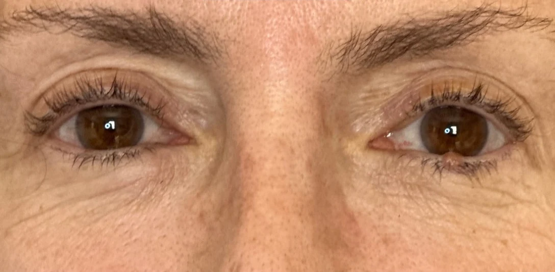
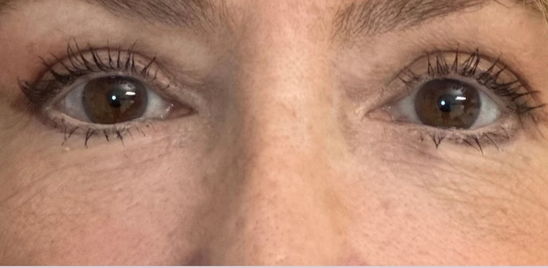
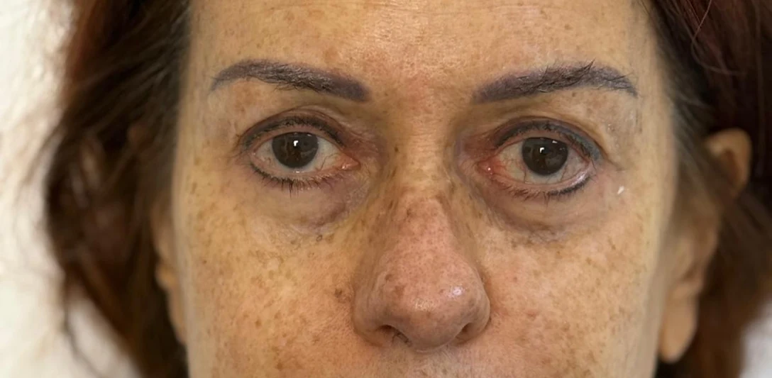
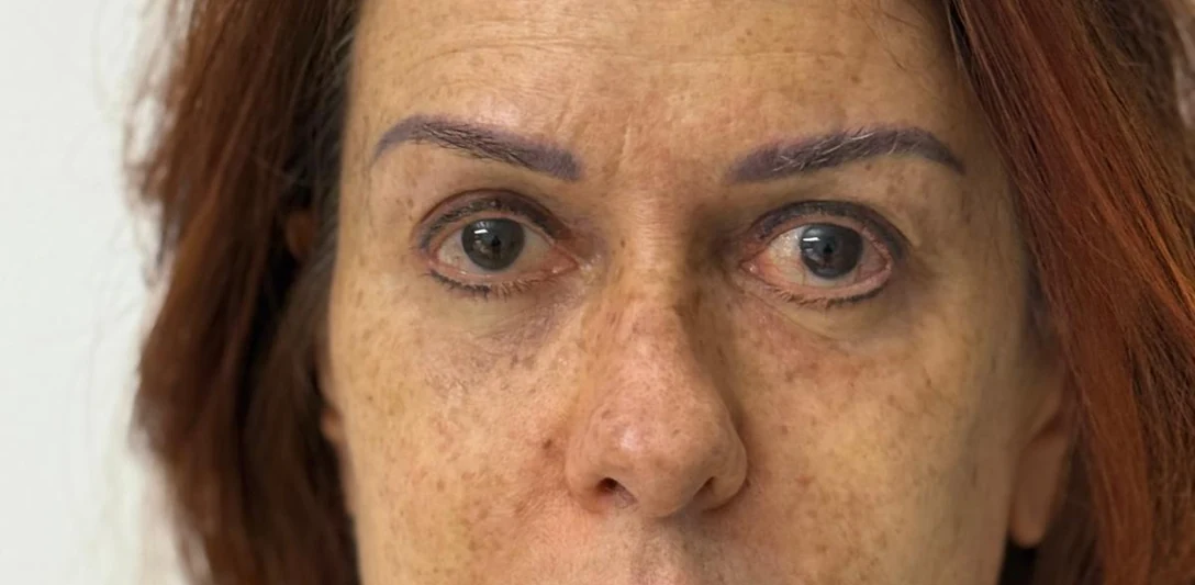
Real cases from the practice, unretouched, framed on the treated area only. Each result is individual and does not predict yours.
Before
After
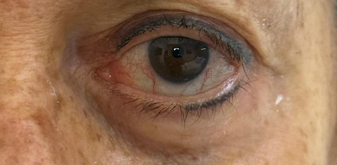
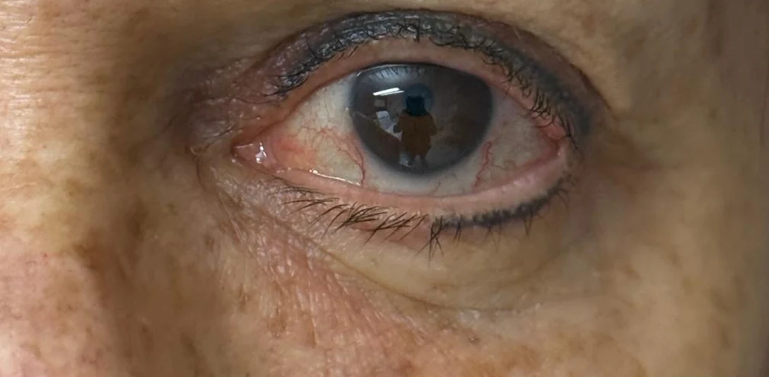
Correcting a hollowed eye area
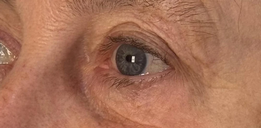
Before
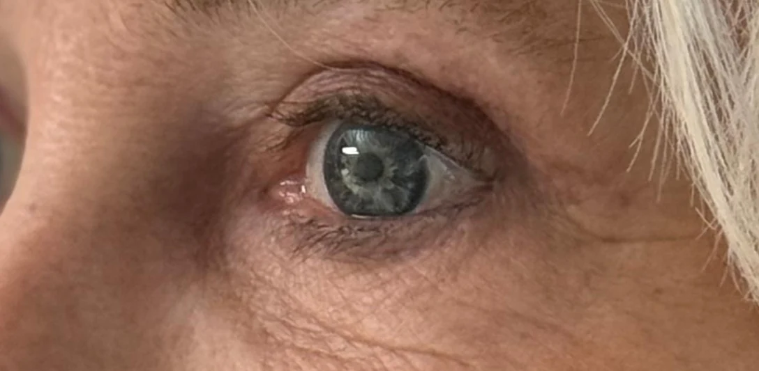
After
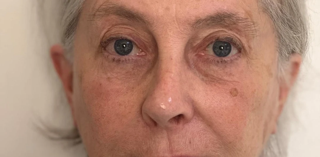
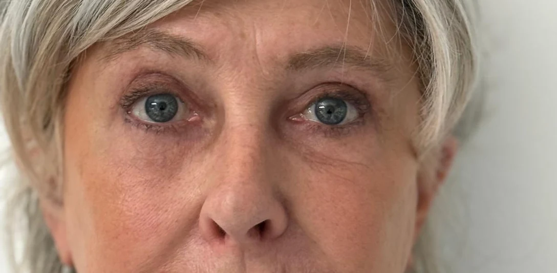
Correcting a hollowed eye area (second case)
Photographs published with the patients’ written consent.
The consequences of an over-aggressive blepharoplasty bear precise names: hollow eye, round eye, ectropion, lagophthalmos, dry eye. Knowing them before undergoing surgery means understanding why the dosage and the surgical approach count as much as the procedure itself — and why the preoperative assessment of the orbital vector, eyelid laxity and tear film weighs as heavily as the surgeon’s hand.
Dr Bernard Hayot · ophthalmologist, former chief resident, trained in oculoplastic surgery. Paris 8e · RPPS 10003926226.
Hollow eye after a first procedure · case treated in December 2024
The hollow eye results from several combined excesses. At the upper eyelid, too much skin and muscle was removed without taking account of the fat loss specific to this region. At the lower eyelids, the removal of the fat pads was too aggressive, and no account was taken of the treatment of the hollow tear trough either.
What can be done
This is one of the main indications for restoration by lipostructure: giving back the volume that has been removed.
The round eye occurs above all after classic blepharoplasties, that is to say by the anterior approach at the level of the lower eyelid. It manifests itself as scleral show: the appearance of white (sclera) visible between the cornea and the lower eyelid in the primary position of gaze.
What can be done
What can still be done depends on the examination: how long ago the procedure was performed, the state of eyelid closure, the quality of the tear film, the tone of the canthal tendon. Not all of these situations can be corrected.
Ectropion represents the extreme case of complication after blepharoplasty: the eyelid is everted, turned outwards, and no longer touches the eyeball. It thus loses its protective function, which leads to numerous ophthalmological symptoms: watering, irritation.
What can be done
What can still be done depends on the examination: how long ago the procedure was performed, the state of eyelid closure, the quality of the tear film, the tone of the canthal tendon. Not all of these situations can be corrected.
Lagophthalmos is the impossibility of closing the eye completely, occurring above all at night..
What can be done
What can still be done depends on the examination: how long ago the procedure was performed, the state of eyelid closure, the quality of the tear film, the tone of the canthal tendon. Not all of these situations can be corrected.
Dry eye after blepharoplasty occurs above all after an upper blepharoplasty, where a pre-existing dry eye syndrome was present and was not compensated for by appropriate treatment postoperatively.
What can be done
What can still be done depends on the examination: how long ago the procedure was performed, the state of eyelid closure, the quality of the tear film, the tone of the canthal tendon. Not all of these situations can be corrected.
Contrary to a widespread idea, the malar bag is not a pad of fat under the eyes. It is a swelling situated lower down, over the malar bone, and of a different nature: not fat, but a lymphatic oedema linked to slowed drainage. This distinction is essential, since the malar bag is not treated in the same way at all as a fatty eyelid pad.
What can be done
One must know how to retighten the orbicularis muscle by a submuscular lift, and to combine this with a micro-lipostructure which will surround the malar bag in order to conceal it.
If you have already been operated on
The operative report of the first procedure, if it was given to you.
The date of that procedure.
Photographs of your eyes before that first procedure.
These situations are described for information. They cannot all be corrected, and every indication is judged on an examination.
Other corrected eye areas
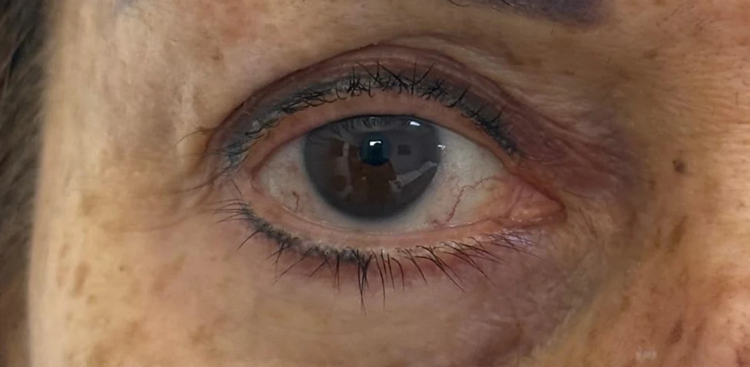
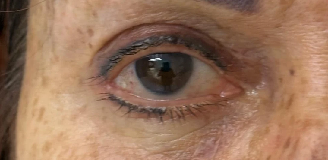
Over-aggressive blepharoplasties, corrected by Dr Hayot. Photographs from the practice, before and after the correction.
Before
After
Correcting a hollowed eye area (third case) · front view
Before
After
Correcting a hollowed eye area (third case) · right eye
Before
After
Correcting a hollowed eye area (third case) · left eye
Before
After
Correcting a hollowed eye area (second case) · front view
1 / 4
Photographs published with the patients’ written consent.
Dr Hayot also explains on Instagram why the eyes can become hollow after a blepharoplasty: view the publication →
Benchmarks from the literature
What the studies say
A few figures published in peer-reviewed journals, chosen to give an idea of the orders of magnitude. They do not describe your case: only the consultation does that.
In a retrospective review covering 892 blepharoplasties performed by a single surgeon over ten years, symptoms of ocular dryness were reported in 26.5 % of patients and chemosis in 26.3 %. The risk was higher when upper and lower blepharoplasty were performed at the same time, and in the case of a transcutaneous approach passing through the orbicularis muscle.
In a series of 50 patients operated on for a lower blepharoplasty by the transcutaneous approach with injection of fat micrografts, five presented a mild ectropion, which resolved without further treatment. No case of infection, of haematoma or of eyelid retraction was observed.
In a survey covering 269,433 cosmetic blepharoplasties reported by oculoplastic surgeons, the incidence of orbital haemorrhage was 0.055 % (about 1 case in 2,000) and that of an orbital haemorrhage with permanent visual loss 0.0045 % (about 1 case in 22,000); the majority of episodes occurred within the first three hours and the risk decreased markedly after twenty-four hours.
The anatomical factors predisposing to lower eyelid retraction after blepharoplasty are a negative orbital vector, horizontal eyelid laxity and a pre-existing retraction; postoperative retraction is related to the failure to treat the laxity, to fibrosis of the middle lamella and to excessive skin resection.
In a series of 1,047 lower blepharoplasties, the incidence of persistent grade 3 conjunctival chemosis was significantly higher after the transcutaneous approach with lateral canthoplasty than after the transconjunctival approach, and higher with the use of radiofrequency.